The Inner World of the Human Cell
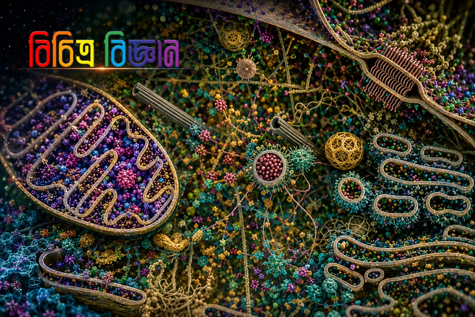
This is neither a digital painting nor a still from a science-fiction film. It is a scientifically accurate visualization of the interior of a human cell—a glimpse into a microscopic realm invisible to the naked eye.
The visualization was assembled from decades of biophysical data, uniting findings from X-ray crystallography, nuclear magnetic resonance (NMR), and cryo-electron microscopy to reconstruct the crowded macromolecular landscape within human biology.
Textbook diagrams often depict cells as tranquil, semi-empty compartments. In reality, a living cell operates like a bustling metropolis. Trillions of protein molecules, ribosomes, mitochondria, and vesicles churn in constant motion: relaying signals, synthesizing ATP, deploying immune defenses, repairing damage, and translating new polypeptide chains every millisecond.
The large purple structure on the left represents the cell nucleus, enclosing chromatin and DNA—the blueprint of the human organism. If the DNA coiled within a single human cell were unraveled and stretched end to end, it would span roughly two meters, yet it sits neatly packed inside a nucleus measuring barely microns across.
Even more staggering: an average human cell contains roughly forty-two million protein molecules, and our bodies are composed of roughly thirty-seven trillion cells. Right now, this vast molecular choreography proceeds silently and continuously inside us, without any conscious direction.
We often marvel at the vastness of the cosmos, yet within our own bodies lies an equally wondrous, teeming molecular universe.