The Mystery of the Cell: Single-Cell Sequencing
The human body is home to roughly 30 trillion cells. Nearly all of them carry identical DNA. Yet, their responsibilities could hardly be more varied. A neuron firing in the brain performs a completely different task than a liver cell, while a white blood cell patrolling the bloodstream operates on an entirely distinct mission. But if they all share the exact same genetic blueprint, why are they so profoundly different?
The secret lies within gene activity. Not all genes are switched on at the same time in every cell. Certain genes are active in some cells, while completely different sets are active in others.
To analyze the genetic profile and gene activity of individual cells in isolation, scientists developed a groundbreaking technique: single-cell sequencing.
Inside the Single Cell
In the past, scientists would bundle thousands of cells together from a tissue sample to analyze their overall gene activity. While this provided a general snapshot, it made it almost impossible to discern what was happening inside individual cells. Single-cell sequencing has transformed this landscape.
Through this technology, researchers can analyze the DNA or RNA of a single cell. Single-cell DNA sequencing identifies subtle genetic changes and mutations within individual cells.
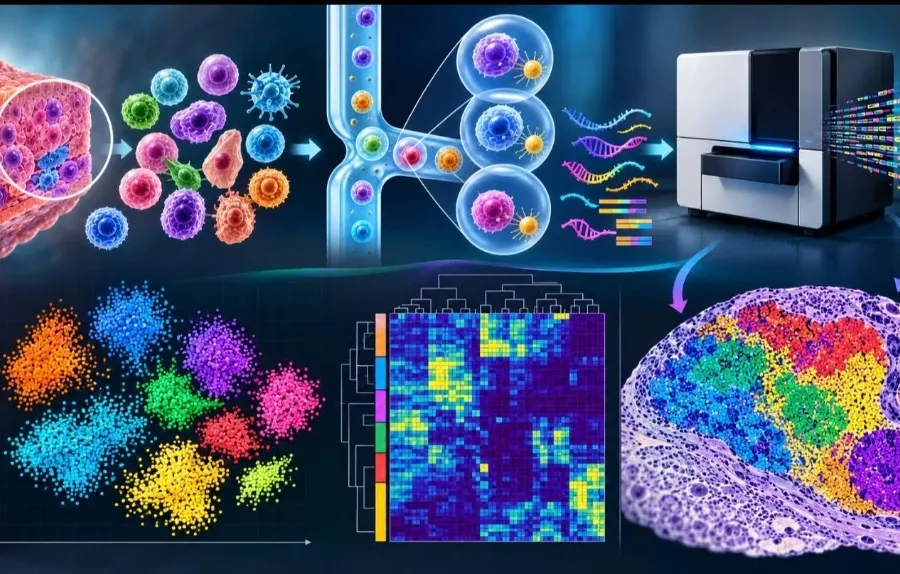
Meanwhile, single-cell RNA sequencing reveals exactly which genes are active. In this approach, individual cells are first isolated from a tissue sample. RNA molecules from each cell are then tagged with unique molecular barcodes. High-throughput sequencing and computational analysis subsequently measure how much RNA is being produced from specific genes in each individual cell. This exposes the fine-grained gene expression of thousands of cells, pinpointing the unique character of every single cell in a crowd.
A Revolution in Cancer Research
Consider a scenario where ten thousand cells are sampled from a patient’s tumor. Under single-cell analysis, it quickly becomes clear that cancer cells are not homogeneous. Some divide aggressively; others express genes linked to drug resistance. Furthermore, the tumor microenvironment harbors diverse immune cells. Previously, bulk tissue analysis concealed these crucial differences in the averages.
Today, researchers can dissect this cellular heterogeneity in unprecedented detail. This opens new frontiers in understanding how tumors proliferate and how they evolve resistance to treatment.
New Horizons in Plant Breeding
The reach of this technology extends far beyond medicine into agricultural and plant research.
Consider a rice plant growing in saline soil. The cells within its roots do not respond uniformly to salt stress. With single-cell techniques, scientists can discover precisely which genes activate in specific cell types within the root. Such insights will be invaluable in breeding salt-tolerant crop varieties.
However, the tough cell walls of plants make isolating intact cells challenging. In many cases, researchers analyze the cell nucleus instead of the entire cell—a variation known as single-nucleus RNA sequencing.
A Step Further: Spatial Transcriptomics
Scientists are now taking this a step further with an exciting complementary technology called spatial transcriptomics.
While single-cell RNA sequencing tells researchers which genes are active within a cell, spatial transcriptomics maps where that cellular activity occurs across the intact tissue.
By pairing both approaches, scientists can uncover not just the identity of a cell, but its physical location and its intricate interactions with neighboring cells.
Biologists once had to settle for bulk impressions of complex tissues. Today, they can look directly inside thousands of individual cells, one by one. Biological discovery has arrived at the single-cell frontier—and that is where the deepest mysteries of life are coming to light.